
Mouse Secondary Antibodies
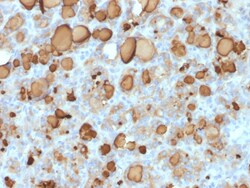
Biotium Thyroglobulin (Thyroidal Cell Marker) (rTGB24), CF740 conjugate, 0.1mg/mL
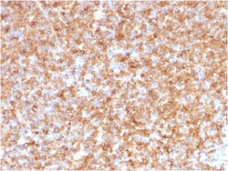
Thyroglobulin is a 660 kDa dimeric pre-protein with multiple glycosylation sites. It is produced by and processed within the thyroid gland to produce the hormone thyroxine and triiodothyronine. Prior to forming dimers, thyroglobulin monomers undergo conformational maturation in the endoplasmic reticulation. The vast majority of follicular carcinomas of the thyroid will give positive immunoreactivity for anti-thyroglobulin even though sometimes only focally. Poorly differentiated carcinomas of the thyroid are frequently anti-thyroglobulin negative. Adenocarcinomas of other-than-thyroid origin do not react with this antibody. This antibody is useful in identification of thyroid carcinoma of the papillary and follicular types. Presence of thyroglobulin in metastatic lesions establishes the thyroid origin of tumor. Anti-thyroglobulin, combined with anti-calcitonin, can identify medullary carcinomas of the thyroid. Furthermore, anti-thyroglobulin, combined with anti-TTF1, can be a


Biotium CELA3B / ELA3B (Pancreatic Function Marker) (CELA3B/1811), CF568 conjugate, 0.1mg/mL
This MAb recognizes a protein of ~30 kDa, identified as CELA3B (Chymotrypsin like elastase family member 3B). Elastases form a subfamily of serine proteases that hydrolyze many proteins in addition to elastin. Humans have six elastase genes which encode the structurally similar proteins elastase 1, 2, 2A, 2B, 3A, and 3B. Unlike other elastases, elastase 3B has little elastolytic activity. Like most of the human elastases, elastase 3B is secreted from the pancreas as a zymogen and, like other serine proteases such as trypsin, chymotrypsin and kallikrein; it has a digestive function in the intestine. Elastase 3B preferentially cleaves proteins after alanine residues. Elastase 3B may also function in the intestinal transport and metabolism of cholesterol. Both elastase 3A and elastase 3B have been referred to as protease E and as elastase 1, and excretion of this protein in fecal material is frequently used as a measure of pancreatic function.Primary antibodies are available pur
Biotium Histone H1 (Nuclear Marker) (r1415-1), Biotin conjugate, 0.1mg/mL
Eukaryotic histones are basic and water-soluble nuclear proteins that form hetero-octameric nucleosome particles by wrapping 146 base pairs of DNA in a left-handed supealpha-helicalturn sequentially to form chromosomal fiber. Two molecules of each of the four core histones (H2A, H2B, H3, and H4) form the octamer; formed of two H2A-H2B dimers and two H3-H4 dimers, forming two nearly symmetrical halves by tertiary structure. Over 80% of nucleosomes contain the linker Histone H1, derived from an intronless gene that interacts with linker DNA between nucleosomes and mediates compaction into higher order chromatin. Histones are subject to posttranslational modification by enzymes primarily on their N-terminal tails, but also in their globular domains. Such modifications include methylation, citrullination, acetylation, phosphorylation, sumoylation, ubiquitination and ADP-ribosylation.Primary antibodies are available purified, or with a selection of fluorescent CF Dyes and other l

Biotium CD4, Mouse (T-Cell Marker)(GK1.5), CF488A conjugate, 0.1mg/mL
CD4 is a member of the Ig superfamily, primarily expressed on most thymocytes, a subset of T cells, and weakly on macrophages and dendritic cells. It acts as a coreceptor with the TCR during T cell activation and thymic differentiation by binding MHC class II and associating with the protein tyrosine kinase, lck. CD4 is expressed by the majority of thymocytes, most helper T cells, a subset of NK-T cells and weakly by dendritic cells and macrophages. CD4 plays an important role in the development of T cells and is required for mature T cells to function optimally. This antibody blocks helper T cell responses to MHC class II antigens, including cytolysis, proliferation, allogeneic B cell help, and release of lymphokines. Primary antibodies are available purified, or with a selection of fluorescent CF Dyes and other labels. CF Dyes offer exceptional brightness and photostability. Note: Conjugates of blue fluorescent dyes like CF405S and CF405M are not recommended for detect
Biotium Wilm's Tumor 1 (WT1) (Wilm's Tumor & Mesothelial Marker), Biotin conjugate, 0.1mg/mL
Recognizes a 47-55 kDa-tumor suppressor protein, identified as Wilm's Tumor (WT1) protein. The antibody reacts with all isoforms of the full-length WT1 and also identifies WT1 lacking exon 2-encoded amino acids, frequently found in subsets of sporadic Wilms tumor and mesothelioma. WT1 protein has been identified in proliferative mesothelial cells, malignant mesothelioma, ovarian carcinoma, gonadoblastoma, nephroblastoma, and desmoplastic small round cell tumor. Lung adenocarcinomas rarely stain positive with this antibody. WT1 protein expression in mesothelial cells has become a reliable marker for the diagnosis of mesotheliomas. Primary antibodies are available purified, or with a selection of fluorescent CF Dyes and other labels. CF Dyes offer exceptional brightness and photostability. Note: Conjugates of blue fluorescent dyes like CF405S and CF405M are not recommended for detecting low abundance targets, because blue dyes have lower fluorescence and can give higher no
Biotium Epstein-Barr Virus (LMP-1)(CS4), CF488A conjugate, 0.1mg/mL
This antibody is a mixture of four different monoclonal antibodies. This antibody is specific to 60 kDa latent membrane protein (LMP-1) encoded by the BNLF1 gene of the EBV. Each clone reacts with different epitopes on the hydrophilic C-terminus of the cytoplasmic domain of LMP-1. This antibody stains strongly with EBV-positive lymphoblastoid cell lines and EBV infected B cell immunoblasts in infectious mononucleosis. EBV, also designated human herpesvirus 4 (HHV-4), is a member of the herpesvirus family and is one of the most common human viruses. EBV infects B cells and, though often asymptomatic, it can cause infectious mononucleosis, a disease characterized by fatigue, fever, sore throat and muscle soreness. Primary antibodies are available purified, or with a selection of fluorescent CF Dyes and other labels. CF Dyes offer exceptional brightness and photostability. Note: Conjugates of blue fluorescent dyes like CF405S and CF405M are not recommended for detecting low
Biotium von Willebrand Factor / Factor VIII Related-Ag (Endothelial Marker) (VWF/1767), CF568 conjugate, 0.1mg/mL
von Willebrand Factor (vWF) is a multimeric glycoprotein that is found in endothelial cells, plasma and platelets. It acts as a carrier protein for Factor VIII and promotes platelet adhesion and aggregation. vWF undergoes a variety of posttranslational modifications that influence the affinity and availability for Factor VIII, including cleavage of the propeptide and formation of N-terminal disulfide bonds. This antibody helps to establish the endothelial nature of some lesions of disputed histogenesis, e.g. Kaposi's sarcoma and cardiac myxoma. It is widely used for differentiating vascular lesions from those of other tissue differentiation within a panel of other vascular markers although not all tumors of endothelial differentiation contain this antigen.Primary antibodies are available purified, or with a selection of fluorescent CF Dyes and other labels. CF Dyes offer exceptional brightness and photostability. Note: Conjugates of blue fluorescent dyes like CF405S and C
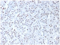
Biotium Nucleophosmin (Acute Myeloid Leukemia Marker)(NPM1/1902), CF568 conjugate, 0.1mg/mL
Recognizes a 33 kDa glycoprotein, identified as Nucleophosmin (NPM). It is predominantly localized in the nucleus of cells in most tissues. NPM is involved in ribosomal assembly and rRNA transport. It is an abundant protein that is highly phosphorylated by Cdc2 kinase during mitosis. This phosphoprotein moves between the nucleus and the cytoplasm. It is thought to be involved in several processes including regulation of the ARF/p53 pathway. A number of genes are fusion partners, in particular the anaplastic lymphoma kinase gene on chromosome 2. Mutations in exon 12 affecting the C-terminus of the protein are associated with an aberrant cytoplasmic location. Mutations in this gene are associated with acute myeloid leukemia. The antibody may be a useful aid for classification of acute myeloid leukemia. Primary antibodies are available purified, or with a selection of fluorescent CF Dyes and other labels. CF Dyes offer exceptional brightness and photostability. Note: Conjugat
Biotium CD30 / TNFRSF8 (Hodgkin & Reed-Sternberg Cell Marker) (rCD30/412), CF568 conjugate, 0.1mg/mL
This antibody recognizes a single chain glycoprotein of 105/120 kDa, identified as CD30/Ki-1. CD30 is synthesized as a 90 kDa precursor, which is processed in the Golgi complex into a membrane-bound phosphorylated mature 105/120 kDa glycoprotein. In Hodgkin's disease, CD30/Ki-1 antigen is expressed by mononuclear-Hodgkin and multinucleated Reed-Sternberg cells. It is also expressed by the tumor cells of a majority of anaplastic large cell lymphomas as well as by a varying proportion of activated T and B cells. This MAb distinguishes large cell lymphomas derived from activated lymphoid cells from histiocytic malignancies and lymphomas derived from resting and precursor lymphoid cells or from anaplastic carcinomas. About one third of the Ki-1 positive lymphomas lack the leukocyte common antigen (CD45).Primary antibodies are available purified, or with a selection of fluorescent CF Dyes and other labels. CF Dyes offer exceptional brightness and photostability. Note: Conjugate
Biotium HLA-DP/-DQ/-DR (MHC II)(CR3/43), CF488A conjugate, 0.1mg/mL
Reacts with a common epitope of human major histocompatibility (MHC) class II antigens, HLA-DP, -DQ and -DR. Human MHC class II antigens are transmembrane glycoproteins composed of an chain (36 kDa) and a chain (27 kDa). They are expressed primarily on antigen presenting cells such as B lymphocytes, monocytes, macrophages, and thymic epithelial cells and are also present on activated T lymphocytes. Human MHC class II genes are located in the HLA-D region that encodes at least six and ten chain genes. Three loci, DR, DQ and DP, encode the major expressed products of the human class II region. The human MHC class II molecules bind intracellularly processed peptides and present them to T-helper cells. They, therefore, have a critical role in the initiation of the immune response. It has been shown that some autoimmune diseases are associated with certain class II alleles.Primary antibodies are available purified, or with a selection of fluorescent CF Dyes and other labels.
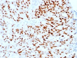
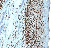
Biotium Estrogen Receptor, alpha (Marker of Estrogen Dependence)(ESR1/1904), 0.2mg/mL
This monoclonal antibody is specific to estrogen receptor alpha (ER alpha) and shows minimal cross-reaction with other members of the family. ER is an important regulator of growth and differentiation in the mammary gland. Presence of ER in breast tumors indicates an increased likelihood of response to anti-estrogen (e. g. tamoxifen) therapy. It strongly stains nuclei of epithelial cells in breast carcinomas. Primary antibodies are available purified, or with a selection of fluorescent CF Dyes and other labels. CF Dyes offer exceptional brightness and photostability. Note: Conjugates of blue fluorescent dyes like CF405S and CF405M are not recommended for detecting low abundance targets, because blue dyes have lower fluorescence and can give higher non-specific background than other dye colors.

Biotium Thyroglobulin (Thyroidal Cell Marker) (rTGB24), CF740 conjugate, 0.1mg/mL
Thyroglobulin is a 660 kDa dimeric pre-protein with multiple glycosylation sites. It is produced by and processed within the thyroid gland to produce the hormone thyroxine and triiodothyronine. Prior to forming dimers, thyroglobulin monomers undergo conformational maturation in the endoplasmic reticulation. The vast majority of follicular carcinomas of the thyroid will give positive immunoreactivity for anti-thyroglobulin even though sometimes only focally. Poorly differentiated carcinomas of the thyroid are frequently anti-thyroglobulin negative. Adenocarcinomas of other-than-thyroid origin do not react with this antibody. This antibody is useful in identification of thyroid carcinoma of the papillary and follicular types. Presence of thyroglobulin in metastatic lesions establishes the thyroid origin of tumor. Anti-thyroglobulin, combined with anti-calcitonin, can identify medullary carcinomas of the thyroid. Furthermore, anti-thyroglobulin, combined with anti-TTF1, can be a
Biotium S100B (Astrocyte and Melanoma Marker) (S100B/1706R), Biotin conjugate, 0.1mg/mL
S100A and S100B proteins are two members of the S100 family of calcium binding proteins. S100A is composed of an alpha and a beta chain whereas S100B is composed of two beta chains. This antibody is specific against an epitope located on the beta-chain (i.e. in S-100A and S-100B) but not on the alpha-chain of S-100 (i.e. in S-100A and S100A0). This antibody can be used to localize S-100A and S-100B in various tissue sections. S-100 protein has been found in normal melanocytes, Langerhans cells, histiocytes, chondrocytes, lipocytes, skeletal and cardiac muscle, Schwann cells, epithelial and myoepithelial cells of the breast, salivary and sweat glands, as well as in glial cells. Neoplasms derived from these cells also express S-100 protein, albeit non-uniformly. A large number of well-differentiated tumors of the salivary gland, adipose and cartilaginous tissue, and Schwann cell-derived tumors express S-100 protein. Almost all malignant melanomas and cases of histiocytosis X ar
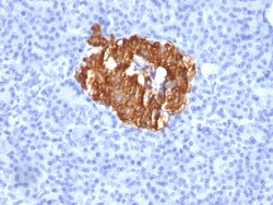
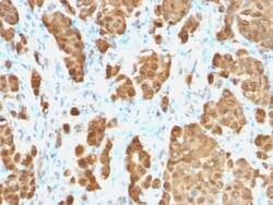
Biotium Insulin / IRDN (beta-Cell & Insulinoma Marker)(K36aC10), Biotin conjugate, 0.1mg/mL
Recognizes a polypeptide which is identified as insulin, a 51-amino acid polypeptide composed of A and B chains connected through the C-peptide. Proinsulin, which has very little biological activity, is cleaved by proteases within its cell of origin into the insulin molecule and the C-terminal basic residue. Insulin enhances membrane transport of glucose, amino acids, and certain ions. It also promotes glycogen storage, formation of triglycerides, and synthesis of proteins and nucleic acids. Deficiency of insulin results in diabetes mellitus. The main storage site for insulin is the pancreatic islets. Antibodies to insulin are important as beta-cell and insulinoma marker. Primary antibodies are available purified, or with a selection of fluorescent CF Dyes and other labels. CF Dyes offer exceptional brightness and photostability. Note: Conjugates of blue fluorescent dyes like CF405S and CF405M are not recommended for detecting low abundance targets, because blue dyes hav
Biotium TTF-1 / NKX2.1 (Thyroid & Lung Epithelial Marker) (rNX2.1/690), Biotin conjugate, 0.1mg/mL
This antibody recognizes a protein of 40 kDa, identified as Thyroid transcription factor-1 (TTF-1). TTF-1 is a member of the NKx2 family of homeodomain transcription factors. It is expressed in epithelial cells of the thyroid gland and the lung. Nuclei from liver, stomach, pancreas, small intestine, colon, kidney, breast, skin, testes, pituitary, prostate, and adrenal glands are unreactive. Anti-TTF-1 is useful in differentiating primary adenocarcinoma of the lung from metastatic carcinomas originating in the breast, mediastinal germ cell tumors, and malignant mesothelioma. It can also be used to differentiate small cell lung carcinoma from lymphoid infiltrates. Loss of TTF-1 expression in non-small cell lung carcinoma has been associated with aggressive behavior of such neoplasms. TTF-1 reactivity is also seen in thyroid malignancies.Primary antibodies are available purified, or with a selection of fluorescent CF Dyes and other labels. CF Dyes offer exceptional brightness